




Melasma is a chronic, relapsing disorder of acquired cutaneous hyperpigmentation presenting as symmetrical, irregular brown-to-slate-gray macules and patches predominantly across the centrofacial, malar, and mandibular regions. Far more than a superficial cosmetic blemish, melasma represents a complex photo-aging and neurovascular dermatosis characterized by biologically hyperactive melanocytes, localized basement membrane disruption, altered dermal elastosis, and vascular endothelial proliferation. The condition is fueled by female steroid hormones (estrogen and progesterone), thermal heat exposure, and ultraviolet as well as high-energy visible (HEV) blue light. Clinical dermatology establishes 7 evidence-based strategies: implementing broad-spectrum tinted mineral photoprotection fortified with iron oxides to neutralize visible light, downregulating melanogenesis through botanical tyrosinase inhibition with aloesin, applying topical azelaic acid and stabilized L-ascorbic acid for competitive enzymic blockade, restoring stratum corneum barrier integrity with colloidal oatmeal and ceramides to prevent subclinical inflammatory cascades, protecting against dermal free radicals with topical green tea epigallocatechin gallate (EGCG), accelerating cellular turnover through the non-irritating retinoid alternative bakuchiol, and stabilizing systemic neuroendocrine triggers through cortisol and estrogen receptor management.
Dermatological Summary & Key Takeaways
- The Iron Oxide Imperative: Standard clear chemical sunscreens only protect against ultraviolet (UVA/UVB) rays; visible blue light (400–500 nm) induces intense, long-lasting hyperpigmentation in skin of color, which can only be physically blocked by tinted sunscreens containing iron oxides.
- Debunking Kitchen Acid Blunders: Applying raw lemon juice, undiluted apple cider vinegar, or crushed horseradish directly to melasma induces chemical burns, phytophotodermatitis, and severe rebound post-inflammatory hyperpigmentation (PIH).
- Multi-Pathway Tyrosinase Blockade: Melanin synthesis cannot be halted by a single ingredient; effective topical management requires combining competitive tyrosinase inhibitors (aloesin, azelaic acid, vitamin C) with barrier-repairing soothing agents.
- The Infrared & Heat Trigger: Intense heat from saunas, hot yoga, and cooking stimulates microvascular dilation and vascular endothelial growth factor (VEGF), aggravating melanocyte output even in total darkness.

Pathophysiology of Melasma: The Melanocyte-Keratinocyte-Fibroblast Axis
Melasma is not merely a problem of excess pigment; it is a multifactorial dermatosis occurring within a senescent, chronically photo-damaged cutaneous microenvironment. Within the epidermal basal layer, hyperfunctional melanocytes synthesize melanin inside lysosome-related organelles called melanosomes. The rate-limiting enzyme in this cascade is tyrosinase, which oxidizes L-tyrosine to 3,4-dihydroxyphenylalanine (L-DOPA) and subsequently into DOPAquinone, ultimately forming dark eumelanin.
In melasma lesions, estrogen receptors (ER-β) and progesterone receptors are markedly upregulated on melanocytes. When stimulated, these receptors trigger downstream cyclic adenosine monophosphate (cAMP) and protein kinase A (PKA) pathways, upregulating microphthalmia-associated transcription factor (MITF)—the master regulator of tyrosinase gene expression. Simultaneously, matrix metalloproteinases (MMP-2 and MMP-9) degrade the type IV collagen scaffolding of the basement membrane, allowing free melanin granules and melanocytes to drop downward into the superficial dermis (dermal melasma), where they are engulfed by melanophages. As detailed in dermatological evaluations published by the National Center for Biotechnology Information (NCBI) on melasma pathogenesis and photoprotection, successful management requires blocking both light-induced enzymatic production and chronic dermal inflammation. For complementary cellular rejuvenation, explore our guide on astaxanthin and marine collagen for dermal defense.
“Melasma is a hyperreactive neurovascular condition where melanocytes act as sensory defense units; aggressive chemical irritation or heat exposure triggers immediate inflammatory pigment defense cascades.”
7 Science-Backed Strategies for Melasma Management
The following 7 evidence-based protocols address melanin synthesis, cellular inflammation, and environmental triggers without provoking secondary post-inflammatory hyperpigmentation:
1. Broad-Spectrum Mineral Photoprotection Fortified with Iron Oxides
Ultraviolet radiation (both UVA and UVB) has long been established as the primary exogenous stimulus for melasma, driving reactive oxygen species (ROS) that stimulate keratinocytes to release stem cell factor (SCF) and endothelin-1. However, groundbreaking photobiological research confirms that high-energy visible (HEV) blue light (400–500 nm) induces intense, long-lasting hyperpigmentation that persists longer than UV-induced erythema in Fitzpatrick skin types III through VI.
Conventional clear chemical and untinted mineral sunscreens filter UV rays but transmit visible blue light almost entirely. Iron oxides (Fe2O3, Fe3O4)—the mineral pigments that tint dermatological foundations and sunscreens—absorb and scatter HEV light across the 400–500 nm spectrum. A randomized, split-face clinical trial demonstrated that applying a broad-spectrum mineral sunscreen (SPF 50+ containing zinc oxide and titanium dioxide) enriched with at least 3% iron oxides significantly diminished melasma relapse rates and Melasma Area and Severity Index (MASI) scores compared to untinted SPF 50+ formulations. Daily reapplication every 2 hours during outdoor exposure is non-negotiable.
2. Botanical Tyrosinase Inhibition via Aloesin (Aloe Vera Bioactive Matrix)
Fresh aloe vera (Aloe barbadensis) contains a pharmacologically active chromone derivative known as aloesin. Unlike harsh chemical bleaches that can cause permanent melanocyte cell death (leukoderma), aloesin acts as a natural, non-toxic, competitive inhibitor of human tyrosinase.
Biochemical assays published by the National Center for Biotechnology Information (NCBI) on aloesin melanogenesis inhibition demonstrate that aloesin binds directly to the active catalytic site of tyrosinase, competitively blocking the conversion of L-tyrosine to L-DOPA. When applied topically in standardized emulsions, aloesin attenuates UV-induced epidermal hyperpigmentation in a dose-dependent manner. Pure, stabilized cold-pressed aloe vera gel also supplies polysaccharides (acemannan) that reduce localized prostaglandin E2 (PGE2) formation, calming cutaneous inflammation and supporting tissue regeneration.
3. Azelaic Acid & Stabilized L-Ascorbic Acid for Competitive Enzyme Blockade
Azelaic acid is a naturally occurring saturated dicarboxylic acid derived from whole-grain cereals and the yeast Malassezia furfur. In clinical dermatology, 15% to 20% topical azelaic acid represents a gold-standard first-line topical treatment for epidermal melasma, demonstrating clinical efficacy comparable to 4% hydroquinone without the perilous risk of exogenous ochronosis (permanent slate-blue skin discoloration).
Azelaic acid acts selectively on abnormal, hyperactive melanocytes, exerting minimal effect on normally functioning surrounding skin. It functions as a competitive inhibitor of tyrosinase while simultaneously suppressing mitochondrial oxidoreductases and inhibiting DNA synthesis in hyperactive melanocytic clones. When paired with stabilized topical vitamin C (10% to 15% L-ascorbic acid formulated at a low pH below 3.5), ascorbic acid acts as an electron donor, reducing DOPAquinone back to DOPA and scavenging light-induced free radicals in the aqueous dermal compartment. Learn proper formulation selection in our guide on how to choose and use vitamin C serum safely.
4. Stratum Corneum Barrier Restoration with Colloidal Oatmeal & Ceramides
Melasma lesions exhibit marked structural defects in stratum corneum barrier integrity, including diminished epidermal ceramide concentrations, elevated transepidermal water loss (TEWL), and persistent low-grade subclinical inflammation. When the lipid matrix is compromised, environmental allergens and UV rays penetrate deeply, activating epidermal toll-like receptors (TLRs) that amplify melanogenesis.
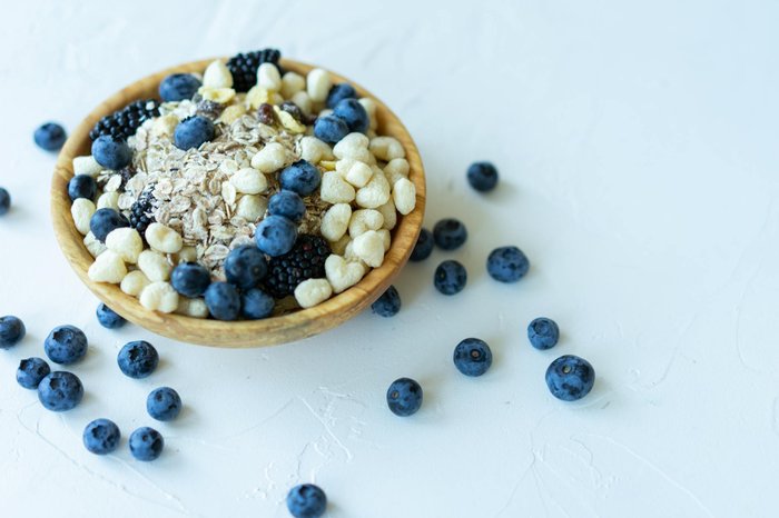
Colloidal oatmeal (Avena sativa) is a standardized botanical preparation rich in beta-glucans, lipids, and specialized phenolic antioxidants termed avenanthramides. Avenanthramides directly inhibit nuclear factor-κB (NF-κB) phosphorylation in keratinocytes, curtailing interleukin-8 (IL-8) and TNF-α release. Incorporating a barrier-repair cream enriched with physiological lipids (3:1:1 ratio of ceramides, cholesterol, and free fatty acids) paired with colloidal oatmeal restores membrane impermeability, shields melanocytes from micro-inflammatory signaling, and prevents irritation from active topicals.
5. Topical & Oral Green Tea Polyphenols (EGCG) and Silymarin
Epigallocatechin gallate (EGCG), the predominant catechin in green tea (Camellia sinensis), and silymarin, a flavonolignan complex extracted from milk thistle (Silybum marianum), represent potent plant bioactives capable of downregulating melanin synthesis through multiple molecular checkpoints.
EGCG downregulates tyrosinase activity not by toxic ablation, but by enhancing the endosomal degradation of mature tyrosinase proteins and reducing intracellular melanin content. Furthermore, topical silymarin has been evaluated in randomized controlled trials against conventional hydroquinone, demonstrating significant drops in MASI scores without cutaneous stinging or erythema. These polyphenols neutralize singlet oxygen (1O2) and lipid peroxides produced during daytime heat and solar exposure, preventing melanocyte membrane peroxidation.
6. Non-Irritating Retinoid Alternatives: Bakuchiol for Cellular Turnover
Traditional topical retinoids (such as prescription tretinoin) accelerate epidermal cell renewal, facilitating the rapid shedding of pigment-laden mature keratinocytes. However, conventional retinoids frequently provoke severe “retinoid dermatitis”—a cascade of peeling, burning, and intense erythema that in darker skin tones (Fitzpatrick types IV–VI) almost inevitably triggers devastating post-inflammatory hyperpigmentation, worsening melasma.
Bakuchiol is a natural meroterpene phenol harvested from the seeds of Psoralea corylifolia. While structurally distinct from vitamin A, bakuchiol operates as a functional retinoid analogue through similar gene expression signatures, stimulating type I collagen synthesis and accelerating basal epidermal turnover without compromising cutaneous barrier lipids or provoking inflammatory skin irritation. In double-blind clinical trials, twice-daily topical application of 0.5% bakuchiol reduced hyperpigmentation surface area and fine photo-aging lines with zero reported retinoid-induced scaling or phototoxicity. Review our comprehensive dermatological overview on bakuchiol as a science-backed natural retinol alternative.
7. Systemic Endocrine & Stress Modulation: Cortisol & Estrogen Management
Melasma is inherently an endocrine-sensitive cutaneous condition, historically designated the “mask of pregnancy” (chloasma). Elevated endogenous estrogen, oral contraceptive pills (OCPs), hormone replacement therapy (HRT), and levonorgestrel intrauterine devices (IUDs) can trigger and maintain melasma recalcitrance.
Furthermore, physiological and psychological stress stimulates the hypothalamic-pituitary-adrenal (HPA) axis, elevating circulating adrenocorticotropic hormone (ACTH) and alpha-melanocyte-stimulating hormone (α-MSH). Both ACTH and α-MSH bind directly to melanocortin 1 receptors (MC1R) on cutaneous melanocytes, igniting rapid intracellular melanin production. Addressing underlying chronic stress through adaptogenic behavioral protocols, prioritizing restorative sleep to regulate circadian cortisol rhythms, and discussing non-hormonal contraception options (such as copper IUDs) with an endocrinologist or gynecologist can remove potent internal neuroendocrine drivers of recurrent melasma.
Medical Debunking: Dangerous Home Remedies That Worsen Melasma
Internet beauty hacks often circulate catastrophic DIY recipes that turn mild epidermal melasma into deep, permanent dermal hyperpigmentation. Strictly avoid these dangerous practices:
- The Raw Lemon Juice Disaster: Applying freshly squeezed lemon juice onto melasma is one of the most destructive DIY skincare trends. Lemon juice has an aggressive acidic pH of approximately 2.0 and contains psoralens (furocoumarins). When exposed to sunlight, psoralens provoke acute phytophotodermatitis—severe blistering and chemical necrosis—followed by dense, intractable post-inflammatory hyperpigmentation.
- Crushed Horseradish & Raw Garlic: Horseradish contains volatile allyl isothiocyanates that act as intense cutaneous irritants. Rubbing raw horseradish onto facial skin destroys epidermal tight junctions, causing chemical burns and stimulating melanocytes to produce defense pigment.
- Undiluted Apple Cider Vinegar: While vinegar contains mild acetic acid, applying undiluted or poorly buffered vinegar strips stratum corneum lipids and causes transepidermal water loss. The resulting chemical erythema triggers inflammatory prostanoids that accelerate tyrosinase transcription.
- Overlooking Environmental Heat (The Thermal Trigger): Many patients assume staying under an umbrella on a hot beach protects their melasma. Ambient thermal heat stimulates transient receptor potential vanilloid channels (TRPV4) and induces cutaneous vasodilation. The resulting influx of inflammatory mast cells and vascular endothelial growth factor (VEGF) activates melanogenesis even without a single direct UV photon reaching the skin.
Comparative Mechanism & Evidence Matrix: Melasma Protocols vs. Dermatological Targets
| Therapeutic Agent / Modality | Molecular Target & Pigment Pathway | Dermatological Evidence & Safety Guidelines |
|---|---|---|
| 1. Tinted Mineral Sunscreen (Iron Oxides) | Absorbs and scatters high-energy visible blue light (400–500 nm) and UVA/UVB | Broad-spectrum SPF 50+ containing ≥ 3% iron oxides; apply daily every 2 hours outdoors |
| 2. Aloesin (Aloe Vera Extract) | Competitive human tyrosinase inhibition; downregulates DOPA synthesis | Safe, non-cytotoxic; apply cold-pressed stabilized gel twice daily; soothing anti-inflammatory |
| 3. Topical Azelaic Acid (15–20%) | Selective inhibition of hyperactive melanocytes; suppresses thioredoxin reductase | First-line clinical efficacy equivalent to hydroquinone without risk of exogenous ochronosis |
| 4. Colloidal Oatmeal & Ceramides | Suppresses NF-κB and IL-8 via avenanthramides; restores 3:1:1 lipid barrier | Reduces transepidermal water loss (TEWL); protects sensitive skin from micro-inflammation |
| 5. Green Tea Polyphenols (EGCG) | Accelerates endosomal tyrosinase degradation; neutralizes singlet oxygen (1O2) | Topical formulations (1–2%) or oral standardized extracts; powerful photoprotective antioxidant |
| 6. Topical Bakuchiol (0.5–1%) | Retinoid-like gene expression; speeds epidermal turnover without retinoid dermatitis | No photosensitization; safe for daytime and nighttime use across all Fitzpatrick skin phototypes |
| 7. Neuroendocrine Modulation | Blunts ACTH, α-MSH, and estrogen receptor-mediated cAMP/PKA melanogenesis | Stress resilience protocols, restorative sleep, and evaluation of non-hormonal contraceptive methods |
Disclaimer: This article is for informational purposes only and is not intended as medical advice. Always consult with a qualified healthcare professional before making changes to your health regimen.
Frequently Asked Questions (FAQ)
Can melasma ever be completely cured permanently?
No, melasma is a chronic, relapsing condition characterized by biologically altered melanocytes that retain cellular memory. While melasma can be successfully cleared and controlled to near invisibility using consistent tyrosinase inhibitors, gentle turnover agents, and strict photoprotection, it cannot be permanently cured. A single afternoon of unprotected sun exposure, intense thermal heat (such as a sauna), or hormonal fluctuations can reactivate resting melanocytes and trigger a recurrence within days.
Why did my melasma get darker after applying lemon juice and apple cider vinegar?
Raw lemon juice contains high concentrations of citric acid and psoralens, which trigger phototoxic chemical burns when exposed to daylight, while vinegar disrupts the epidermal stratum corneum barrier. Because melanocytes in melasma patients are in a state of hyperreactive alert, acute chemical trauma sparks an immediate inflammatory cascade (post-inflammatory hyperpigmentation), forcing melanocytes to deposit dense melanin deeper into the dermal tissue.
Are chemical peels or intense pulsed light (IPL) safe for treating melasma?
Extreme caution is required. Aggressive chemical peels and IPL devices generate substantial cutaneous heat and tissue injury. In Fitzpatrick skin phototypes III through VI, the inflammatory response from IPL or deep peels frequently provokes severe rebound melasma that is more recalcitrant than the original condition. Most board-certified dermatologists prioritize gentle, superficial peels (such as low-strength mandelic, lactic, or azelaic acid) and low-fluence Q-switched lasers only after topical pigment stabilization.