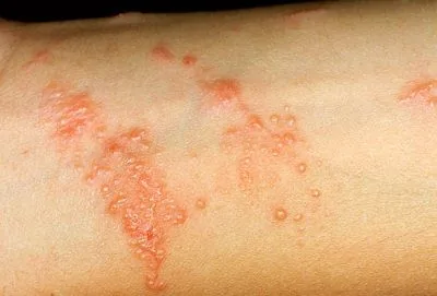
Contact with poison ivy (Toxicodendron radicans) inoculates the epidermal stratum corneum with urushiol—a potent mixture of pentadecacatechol compounds that rapidly penetrate lipid cell membranes. Within 10 to 30 minutes, urushiol oxidizes and irreversibly binds as an allergenic hapten to cutaneous membrane proteins, priming Langerhans cells and triggering an aggressive Type IV cell-mediated delayed hypersensitivity cascade. Subsequent re-exposure unleashes interferon-gamma (IFN-γ) and cytotoxic T lymphocytes, resulting in marked intraepidermal spongiosis, linear vesicular eruptions, severe dermal edema, and agonizing pruritus. While widespread systemic reactions and mucous membrane involvement require immediate clinical corticosteroids, localized poison ivy dermatitis can be rapidly alleviated through 6 clinically supported, evidence-based home interventions: immediate surfactant-solvent decontamination to dissolve unbound urushiol prior to haptenation, colloidal oatmeal soaks to suppress NF-κB signaling via bioactive avenanthramides, alkaline sodium bicarbonate compresses to neutralize pruritic inflammatory acidosis and withdraw exudate, targeted cryotherapy to slow C-fiber action potentials and induce microvascular vasoconstriction, pure cold-pressed aloe vera hydrogel to degrade inflammatory bradykinin via bradykinase, and dilute astringent compresses to precipitate weeping blister proteins and fortify the epidermal acid mantle.
Dermatological Summary & Urgent Anti-Urushiol Protocols
- The Critical Decontamination Window: Unbound urushiol remains accessible to organic solvents and degreasing surfactants for approximately 10 to 30 minutes following contact; thorough emulsification during this latency window dramatically truncates subsequent immune activation.
- Intraepidermal Vesicular Integrity: The serous fluid filling poison ivy bullae consists of host transudate and cellular debris completely devoid of urushiol; preserving intact blister roofs prevents bacterial superinfection by Staphylococcus aureus.
- Neurovascular Pruritus Interruption: Sustained local cooling below 15°C (59°F) physically blunts peripheral C-fiber action potential velocity while constricting postcapillary venules to halt inflammatory serum exudation.
Pathophysiology of Urushiol Contact Dermatitis: Haptenation, Spongiosis, and Pruriceptive Signaling
Poison ivy dermatitis is not an infectious process or an irritant burn; it represents a classical delayed-type (Type IV) cell-mediated hypersensitivity reaction. The resinous sap of the plant contains urushiol, an oleoresin characterized by a catechol ring substituted with a 15- or 17-carbon aliphatic hydrocarbon side chain possessing variable degrees of unsaturation. Due to its intense lipophilicity, urushiol rapidly traverses the cornified stratum corneum.
Once inside the viable epidermis, the catechol ring undergoes enzymatic or auto-oxidation into reactive quinone intermediates. These electrophilic quinones covalently cross-link with self-proteins on epidermal keratinocytes and dendritic cells, forming stable hapten-protein conjugates. Resident antigen-presenting cells—specifically CD1a-expressing epidermal Langerhans cells—internalize these conjugates, migrate along afferent lymphatics to regional draining lymph nodes, and present urushiol peptides on major histocompatibility complex (MHC) class I and class II molecules to naive T lymphocytes. This immunogenic priming generates persistent populations of antigen-specific CD4+ T-helper 1 (Th1) and CD8+ cytotoxic T cells.
Upon subsequent re-exposure, sensitized memory T cells rapidly infiltrate the dermis, releasing a surge of pro-inflammatory cytokines including interferon-gamma (IFN-γ), tumor necrosis factor-alpha (TNF-α), interleukin-17 (IL-17), and interleukin-8 (IL-8). As detailed in comprehensive clinical monographs from the National Institutes of Health (NIH) StatPearls on allergic contact dermatitis and urushiol mechanisms, this immune barrage breaks down intercellular desmosomes between keratinocytes—a pathological process designated as spongiosis. Intercellular edema coalesces into intraepidermal microvesicles, which progressively enlarge into macroscopic weeping bullae. Concurrently, inflammatory neuropeptides and kinins directly sensitize unmyelinated polymodal C-fiber pruriceptors, transmitting fierce itch signals to the dorsal horn of the spinal cord. Implementing rapid, barrier-focused remedies alongside our dermatological guide on home remedies for clear and healthy skin ensures foundational epidermal recovery.
“Urushiol itself ceases to spread across the body once thoroughly bound to epidermal membrane proteins; the appearance of new blisters days later merely reflects regional variations in skin permeability, stratum corneum thickness, and initial antigen dosage.”
6 Science-Backed Home Remedies for Poison Ivy
Managing acute poison ivy dermatitis requires dual therapeutic priorities: terminating unabsorbed antigen exposure during the golden window and blunting the cutaneous inflammatory cascade once the hypersensitivity reaction initiates. The following 6 interventions provide robust, scientifically documented relief:
1. Early Surfactant & Solvent Decontamination (Pre-Haptenation Urushiol Stripping)
The single most decisive intervention in poison ivy management is mechanical and chemical decontamination performed immediately following suspected contact. Because urushiol is a non-polar, hydrophobic lipid resin, plain cold water alone is completely incapable of solubilizing the oil; water molecules roll off the resinous film, permitting ongoing trans-stratum corneum diffusion.
To successfully neutralize the allergen, one must introduce an organic solvent or a potent degreasing amphiphilic surfactant within the first 10 to 30 minutes of exposure. Applying isopropyl alcohol (rubbing alcohol, 70%) or heavy-duty degreasing liquid dishwashing detergent efficiently partitions the non-polar pentadecacatechol chains into micellar structures, lifting the oil away from the epidermal lipid mantle. Wash the skin vigorously using a washcloth, moving in a unidirectional wiping pattern to avoid redistributing the allergen over unexposed cutaneous territory, and rinse thoroughly with copious volumes of lukewarm or cool water. Thoroughly laundering contaminated garments, shoes, and tools is equally critical, as anhydrous urushiol remains chemically active and capable of provoking severe dermatitis for months to years if left unwashed.
2. Colloidal Oatmeal Soaks & Compresses (Avenanthramide-Mediated NF-κB Suppression)
Colloidal oatmeal—pulverized whole oat kernels (Avena sativa) containing finely dispersed starches, beta-glucans, and phytosterols—is an FDA-recognized dermatological protectant that dramatically halts the inflammatory amplification loop of allergic contact dermatitis. Its profound antipruritic potency originates in a rare family of polyphenolic alkaloids found exclusively in oats, known as avenanthramides (principally avenanthramides A, B, and C).
As documented in peer-reviewed dermatological investigations indexed in the National Center for Biotechnology Information (NCBI) on the cutaneous pharmacology of oat avenanthramides, these bioactive phytochemicals inhibit the phosphorylation of the IκB kinase complex. This stops the nuclear translocation of nuclear factor-kappa B (NF-κB) within cutaneous keratinocytes and dermal vascular endothelial cells, resulting in steep reductions in IL-8, TNF-α, and cell adhesion molecules. Furthermore, oat polysaccharides form a hydrophilic hydrocolloid barrier across weeping vesicular lesions, binding excess exudate while sealing moisture into damaged stratum corneum. Soak in a lukewarm bath infused with 1 cup of ultra-fine colloidal oatmeal for 15 to 20 minutes, or blend 2 tablespoons of oatmeal powder with cool water to construct a topical soothing paste for localized rash plaques.
3. Alkaline Sodium Bicarbonate Compresses (Interrupted Pruriceptive Firing & Exudate Osmosis)
Sodium bicarbonate (baking soda, NaHCO3) is an amphoteric mineral salt whose therapeutic utility in poison ivy arises from localized pH buffering and trans-epidermal osmotic fluid shifts. Acute contact dermatitis generates marked interstitial acidosis within the superficial dermis due to cellular necrosis, lactic acid accumulation, and the release of acidic lysosomal hydrolases from invading granulocytes. This acidic microenvironment directly sensitizes acid-sensing ion channels (ASIC) and transient receptor potential vanilloid 1 (TRPV1) channels on cutaneous C-fibers, triggering intractable pruritus.
Applying an alkaline paste of sodium bicarbonate (formulated at a 3:1 ratio of baking soda to filtered water) provides bicarbonate anions (HCO3−) that neutralize superficial cutaneous acids, restoring dermal physiological pH balance. In addition, the hypertonic crystalline paste creates an osmotic concentration gradient across micro-fissured epidermis, drawing stagnant serous transudate out of swollen, weeping vesicles and promoting accelerated lesion drying. Spread the fresh paste gently over itchy plaques for 10 to 15 minutes, allowing it to dry before rinsing with cool water. Combining sodium bicarbonate compresses with gentle itch relief strategies outlined in our review of evidence-based home remedies for mosquito bites establishes comprehensive dermatological relief.
4. Targeted Cold Compresses & Cryotherapy (Postcapillary Venular Vasoconstriction & Conduction Deceleration)
Thermodynamic cooling constitutes the most immediate physiological tool for blunting severe neurogenic itch and throbbing vascular tension. When cutaneous tissue is exposed to icy temperatures, local physiological adaptations occur across both neurological and microvascular compartments.
Physiologically, reducing regional dermal temperatures below 15°C (59°F) substantially reduces the action potential conduction velocity of unmyelinated type C sensory fibers. Simultaneously, cooling activates transient receptor potential melastatin 8 (TRPM8) receptors on cold-sensitive A-delta fibers, sending afferent inhibitory signals to the substantia gelatinosa of the dorsal horn that effectively gate out ascending pruriceptive transmission. On the microvascular level, cold activates local alpha-adrenergic sympathetic pathways, producing profound vasoconstriction of dermal postcapillary venules. This restriction drastically reduces intravascular hydrostatic pressure, preventing further extravasation of serum and reducing tissue edema. Apply a clean washcloth soaked in ice-cold physiological saline or a wrapped ice pack to the eruption for 15 to 20 minutes every 2 to 3 hours. Never apply uncovered ice directly to denuded, blistered skin to avert cryo-induced epidermal necrosis.
5. Pure Cold-Pressed Aloe Vera Hydrogel (Bradykinase-Mediated Peptide Cleavage & Rehydration)
Fresh gel derived from the inner parenchymal tissue of Aloe barbadensis Miller delivers targeted anti-inflammatory and tissue-repairing biochemicals that accelerate re-epithelialization in allergic dermatoses. The primary bioactive structural component of the gel is acemannan, a long-chain beta-1,4-acetylated polymannose polymer that forms a breathable, protective hydrogel shield over excoriated skin.
Crucially for poison ivy relief, aloe vera contains the specialized proteolytic enzyme bradykinase. In the inflamed dermis, cellular injury generates high local concentrations of bradykinin—a vasoactive nonapeptide that induces extreme microvascular permeability, tissue swelling, and pain sensitization. Bradykinase enzymatically cleaves and inactivates bradykinin, curtailing vascular leakage and settling dermal tension. Concurrently, plant sterols (lupeol, campesterol, and beta-sitosterol) suppress cyclooxygenase-2 (COX-2), arresting downstream prostaglandin E2 (PGE2) formation. Apply 100% pure, refrigerated aloe vera gel liberally to the rash 3 to 4 times daily. Confirm that commercial preparations are completely free of drying denatured alcohols or synthetic fragrances, which can provoke secondary irritant dermatitis.
6. Mild Astringent Compresses (Witch Hazel & Dilute Acetic Acid for Blister Weeping)
Once poison ivy progresses to the vesicular and weeping stage, astringent therapy becomes vital to desiccate oozing lesions and fortify cutaneous barrier defenses. Astringents are chemical agents that mildly precipitate surface proteins, condensing and contracting superficial tissue.
Distilled witch hazel (Hamamelis virginiana), rich in gallotannins and hamamelitannin, cross-links surface keratin molecules in exposed bullae, forming a dry, protective coagulum over weeping fissures. Similarly, organic apple cider vinegar diluted 1:2 with sterile water supplies mild acetic acid (CH3COOH) that lowers cutaneous pH back toward its physiological acidic baseline (pH 4.5 to 5.5). This re-acidification stabilizes the epidermal acid mantle, creating an inhospitable terrain for opportunistic bacterial invaders like Staphylococcus aureus. Moisten sterile gauze pads with cold witch hazel or dilute vinegar solution and lay them gently over oozing areas for 10 to 15 minutes twice daily. To review the safe handling and dilution metrics of organic acids, explore our guide on evidence-based apple cider vinegar benefits and safe dilution rules.
Comparative Mechanistic Matrix of Poison Ivy Interventions
| Remedy / Bioactive Agent | Biochemical Target & Physiological Mechanism | Evidence Level | Clinical Protocol & Safety Boundary |
|---|---|---|---|
| 1. Early Surfactant Decontamination | Solubilizes non-polar pentadecacatechol lipid chains into micelles; prevents hapten-protein conjugation. | High (Preventative Standard) | Apply 70% isopropyl alcohol or dish soap within 10–30 minutes of contact. Rinse unidirectionally. |
| 2. Colloidal Oatmeal (Avenanthramides) | Inhibits IκB kinase phosphorylation; blocks NF-κB nuclear translocation; suppresses IL-8 and TNF-α. | High (FDA Protectant) | Soak in lukewarm oat bath for 15–20 minutes or apply thick paste. Pat dry without rubbing. |
| 3. Sodium Bicarbonate Paste (Baking Soda) | Neutralizes dermal metabolic acidosis; desensitizes ASIC/TRPV1 channels; osmotic transudate drying. | Moderate (Empirical / Biochemical) | Mix 3:1 powder to water. Apply for 10–15 minutes maximum, then rinse clean with cool water. |
| 4. Targeted Cold Compresses (Cryotherapy) | Decelerates unmyelinated C-fiber conduction velocity; TRPM8 sensory gating; postcapillary venular constriction. | High (Physiological Standard) | Apply cloth-wrapped cold pack for 15–20 minutes every 2–3 hours. Never apply bare ice to open bullae. |
| 5. Cold-Pressed Aloe Vera Gel | Acemannan hydrogel moisture barrier; bradykinase enzymatic cleavage of bradykinin; COX-2 down-regulation. | Moderate to High | Apply pure chilled gel 3–4 times daily. Ensure zero added alcohol or synthetic dyes. |
| 6. Mild Astringent Compresses (Witch Hazel) | Tannin-mediated protein cross-linking; seals weeping vesicle exudates; re-acidifies the stratum corneum mantle. | Moderate | Dab sterile gauze soaked in witch hazel or 1:2 diluted ACV for 10–15 minutes twice daily. |
Folk Myths, Dangerous Practices & Critical Medical Redlines
Widespread Internet lore regarding poison ivy contains hazardous misconceptions that can exacerbate dermal destruction and ignite life-threatening systemic complications:
- The Scalding Water Fallacy: Running intensely hot water over a poison ivy rash temporarily causes an explosive release of neuronal histamine, yielding a transient sensation of euphoric relief followed by total sensory exhaustion. However, scalding heat inflicts thermal microvascular dilation, dramatically accelerates inflammatory vascular permeability, and worsens dermal spongiosis. Always utilize cool or tepid water.
- The Bleach and Harsh Chemical Hazard: Applying household sodium hypochlorite (bleach) or concentrated coffee concoctions chemically corrodes the already compromised stratum corneum, inflicting chemical burns, severe contact necrosis, and facilitating secondary invasive bacterial cellulitis.
- Blister De-roofing (Excoriation): Never scratch, puncture, or excise the roofs of poison ivy vesicles. The blister roof acts as a sterile biological dressing. Exposing the raw dermal bed creates a direct portal for Staphylococcus aureus and Streptococcus pyogenes superinfections.
- Critical Respiratory Warning: Never burn poison ivy, poison oak, or poison sumac brush. Urushiol does not break down upon combustion; it volatilizes into hot particulate smoke. Inhaling urushiol-laden smoke provokes severe pulmonary mucosal edema, extensive tracheobronchial sloughing, and fatal acute respiratory failure requiring immediate emergency intubation.
- Urgent Clinical Red Flags: Seek immediate medical care if the rash involves the eyelids, periorbital tissue, mouth, or genitalia; if the rash covers greater than 25% of the total body surface area; if streaks of redness, purulent yellow exudate, or systemic fevers develop; or if dyspnea occurs.
Disclaimer: This article is for informational purposes only and is not intended as medical advice. Always consult with a qualified healthcare professional before making changes to your health regimen.
Frequently Asked Questions (FAQ)
Can the fluid inside poison ivy blisters spread the rash to other body parts or people?
No. Poison ivy blister fluid consists purely of host inflammatory serum exudate and cellular transudate; it contains zero urushiol. The rash cannot be transmitted to other skin areas or other individuals through weeping blisters. New lesions appearing days later stem from differential epidermal thickness and varying initial doses of plant oil absorbed during the initial exposure event.
How long after exposure to poison ivy can urushiol be washed off effectively?
Urushiol must be removed within 10 to 30 minutes following contact for maximum preventative benefit. After approximately 30 minutes, the non-polar oleoresin binds irreversibly to epidermal membrane proteins, initiating the immune cascade. However, washing with degreasing surfactants even hours later remains worthwhile to remove unbound surface oil and prevent accidental secondary contamination of other body parts.
When does poison ivy dermatitis require prescription oral corticosteroids?
Prescription systemic corticosteroids (such as an oral prednisone taper lasting 14 to 21 days) are mandatory when the eruption covers more than 20% to 25% of the body surface area, when facial or periorbital edema threatens eye closure, or when lesions involve the mouth or genitalia. Short 5-day steroid bursts must be avoided, as premature discontinuation frequently triggers severe rebound dermatitis before the cell-mediated hypersensitivity reaction has naturally resolved.